Journal of Neurology Research, ISSN 1923-2845 print, 1923-2853 online, Open Access
Article copyright, the authors; Journal compilation copyright, J Neurol Res and Elmer Press Inc
Journal website http://www.neurores.org

Case Report

Volume 1, Number 3, August 2011, pages 121-123


Brain MRI Depiction of Multiple Cranial Neuropathies in Neurosarcoidosis: A Case Report

Figures

Figure 1.
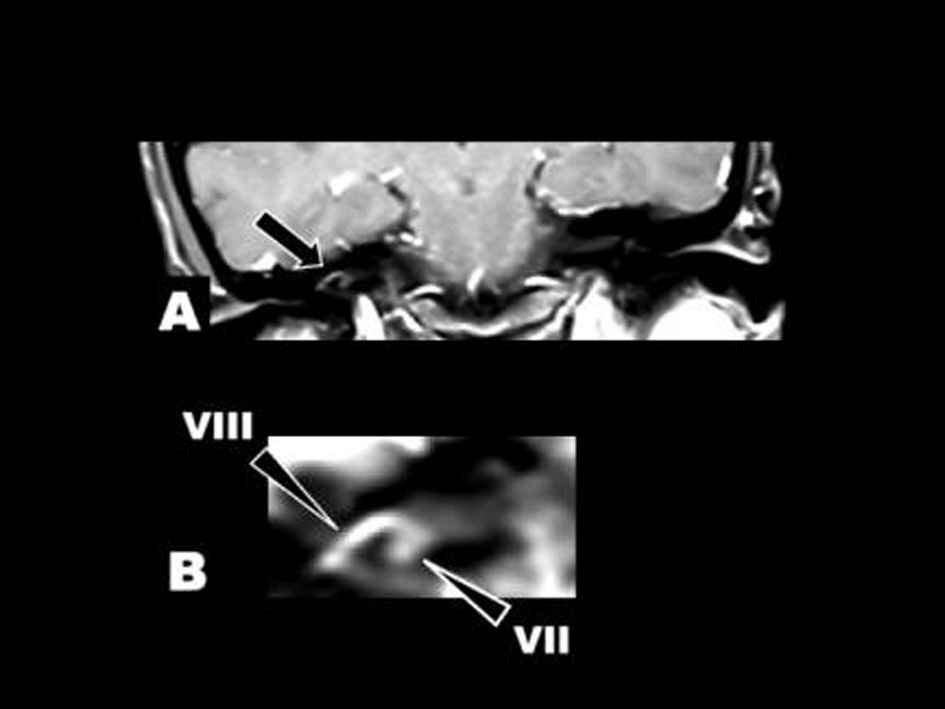
Figure 1. Post-gadolinium T1-weighted imaging at onset of the right VII and VIII cranial neuropathies. (A) Coronal view of the internal auditory canals. Abnormal enhancement was found in the right geniculate ganglion (VII) and the right VIII cranial nerve (arrow). (B) Enlarged view of the right internal auditory canal. Arrowheads indicated VII (the facial nerve) and VIII (the vestibular nerve).
Figure 2.
Figure 2. (A) Ophthalmoscopy showed edema in left optic discs. (B) Severe degree of the left mydriasis. (C) Laryngoscopy showed difficulty in elevating the left pharyngeal wall.
Figure 3.
Figure 3. Post-gadolinium T1-weighted imaging at onset of the left IX and X cranial neuropathies. (A) Axial and (B) coronal view. Abnormal enhancement was found in the bilateral IX and X cranial nerves (arrows). (C) Coronal and (D) sagittal view. Leptomeningeal enhancement was found in the pons and the fourth ventricle (arrowheads).