Journal of Neurology Research, ISSN 1923-2845 print, 1923-2853 online, Open Access
Article copyright, the authors; Journal compilation copyright, J Neurol Res and Elmer Press Inc
Journal website https://www.neurores.org

Case Report

Volume 11, Number 5, October 2021, pages 102-107


Simultaneous Encephalitis and Neuroretinitis After COVID-19 in a Young Adult: A Case Report

Figure

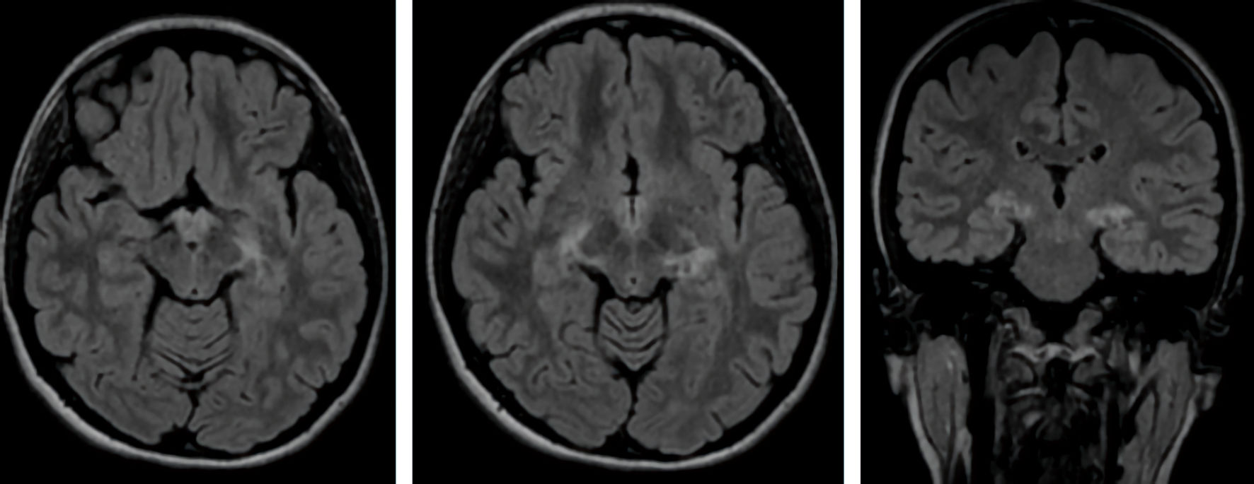
Figure 1.
Figure 1. Axial and coronal views of brain MRI show FLAIR increased signal intensity in the cortical and subcortical regions of both mesial temporal lobe and both side hippocampal tails with a relatively symmetrical appearance. MRI: magnetic resonance imaging; FLAIR: fluid-attenuated inversion recovery.

Table

Table 1. Clinical Presentations of Retinal Involvement After COVID-19
 
No.AuthorAge/sex of the patientClinical presentationsDiagnosisOutcome
Similar presentation (bilateral involvement) was seen in three cases; in most of these patients significant improvement was seen after treatment (similar as our patient). COVID-19: coronavirus disease 2019; M: male; F: female; N/A: not applicable.
1Hosseini et al, 2021 [24]37/MBilateral severe vision lossBilateral retinitis and panuveitisSignificant improvement after treatment
2Goyal et al, 2021 [25]32/MBilateral paracentral and triangular negative scotomaAcute macular neuroretinopathy (AMN) and paracentral acute middle maculopathy (PAMM)No change in vision
3Liu et al, 2021 [23]Elderly/FMonocular blindnessRetinitis and optic neuritisN/A
4Mahendradas et al, 2021 [26]49/FSudden-onset painless loss of vision in both the eyesBilateral post fever retinitis with retinal vascular occlusionsImprovement in vision after treatment
5Our patient18/FBilateral severe vision lossBilateral neuroretinitisSignificant improvement after treatment