Journal of Neurology Research, ISSN 1923-2845 print, 1923-2853 online, Open Access
Article copyright, the authors; Journal compilation copyright, J Neurol Res and Elmer Press Inc
Journal website https://www.neurores.org

Case Report

Volume 000, Number 000, March 2024, pages 000-000


Motor Neuron Disease-Frontotemporal Dementia Spectrum Disorder: A Different Phenotype Related With a Novel TBK1 Gene Variant

Figures

Figure 1.
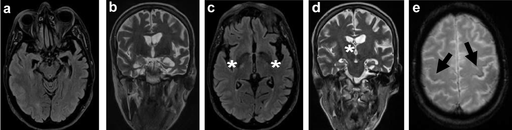
Figure 1. Brain MRI: axial FLAIR (a) and T2-weighted imaging (b) showing asymmetrical temporal lobe atrophy more pronounced on the left. Axial FLAIR (c) and coronal T2-weighted imaging (d) revealing hyperintensity in the posterior limb of the internal capsule and corticospinal tracts (white *). Axial T2 GRE (e) showing iron deposition (loss of signal) in the premotor cortex, most notably in the left precentral gyrus, known as the “motor band sign” (black arrows). MRI: magnetic resonance imaging; FLAIR: fluid-attenuated inversion recovery.
Figure 2.
Figure 2. FDG-PET showing reduced [18F]FDG uptake in the left parietal-temporal and frontal lobes (white *). FDG-PET: fluorodeoxyglucose-positron emission tomography.